FIGURE 2.

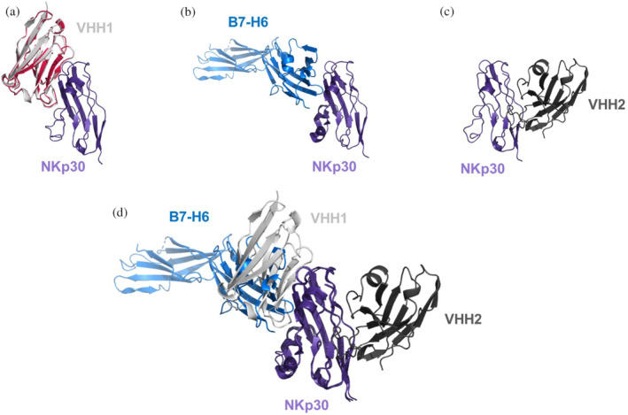
Crystal structures and AlphaFold2 generated binding models of NKp30 with different ligands. (a) X‐ray structure of apo‐VHH1 (shown in red, PDB code 9FXF), superimposed in the AlphaFold2 generated VHH1‐NKp30 complex, reveals that the VHH1 apo structure matches the VHH1 conformation of the AlpaFold2 generated VHH1‐NKp30 complex. (b) Binding mode of the B7‐H6–NKp30 complex (PDB code 3PV6). (c) X‐ray structure of the VHH2‐NKp30 complex (PDB code 9FWW). (d) Superimposition of VHH1‐, VHH2‐, and B7‐H6‐binding modes against NKp30 reveals that the epitopes of B7‐H6 and VHH1 are overlapping, while VHH2 is binding to a distinct epitope.
