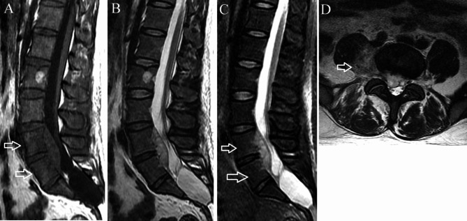
Fig. 1.
Phlegmonous stage primary L4-5 spinal epidural abscess in a 37-year-old man. (A) Sagittal T1-weighted imaging shows a fusiform homogeneously isointense lesion ventral to the thecal sac at L4-5. Edema is present in the L4 and L5 vertebral bodies (arrows). (B) On sagittal T2-weighted imaging, the lesion appears homogeneously hyperintense. (C) On sagittal short tau inversion recovery imaging, the vertebral body edema appears mildly hyperintense (arrows). (D) Axial T2-weighted imaging demonstrates the lesion extending through the neural foramina on the right into the paraspinal region (arrow)

