
Figure 3.
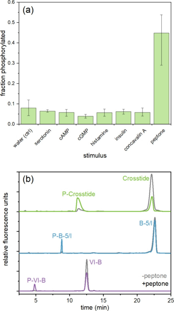
Phosphorylation of peptide substrate reporters in T. thermophila lysates. (a) Average fraction of Crosstide that was phosphorylated after 15 min incubation in lysates prepared 20 s after stimulation with 1 μM stimulus (or 2% for proteose peptone). Error bars represent the maximum and minimum values for n = 2–3 biological replicates on different days. (b) Electropherograms of the three peptide substrate reporters (Crosstide, green; B-5/I, blue; VI–B, purple) incubated with T. thermophila lysates from cells 2 h into conjugation for unstimulated (gray) and stimulated (color) cells with 2% proteose peptone. Cells were lysed just before or 30 s after stimulation. For all traces, x-axes have been normalized to align peaks for display only.