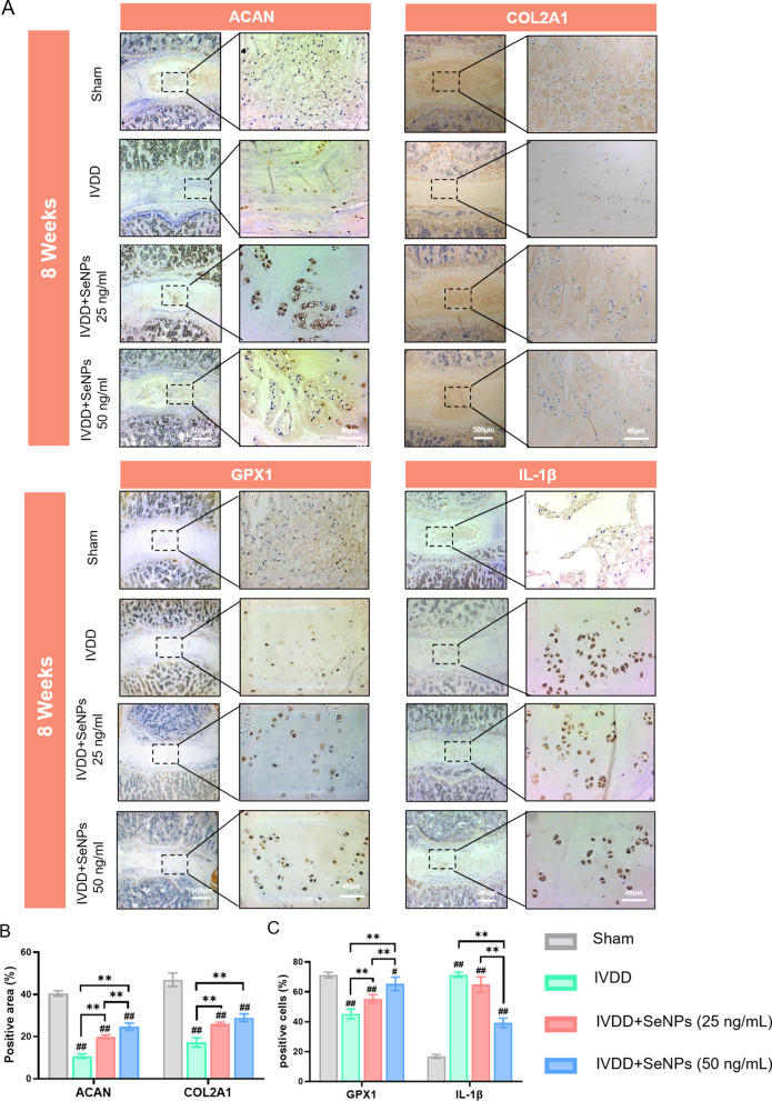
Fig. 10.
Immunohistochemical (IHC) stainings of nucleus pulposus tissues in rats at 4 and 8 weeks after treatment. A Immunohistochemical staining of protein ACAN, COLIIA1, GPX1 and IL-1β of rats’ IVDs at 8 weeks after treatment. B Quantification of the percentages of ACAN-positive and COL2A1-positive area in nucleus pulposus tissue. C Quantification of the percentages of GPX1-positive and IL-1β-positive cells in nucleus pulposus tissue. Statistically significant differences are indicated by # where P < 0.05 or ## where P < 0.01 compared with the control group; * where P < 0.05 or ** where P < 0.01 between the indicated group

