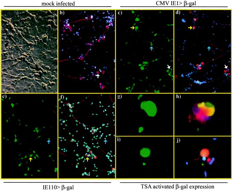
FIG. 2.
Representative examples of mock-infected neuronal cultures or cultures infected with 106 PFU of CS5 (CMV IE1>β-gal) or gH−TK−110LacZ (IE110>β-gal) per well. Cultures were fixed and dually immunostained for expression of β-gal (FITC, green) and neuron-specific β-tub (Cy3, red) and were counterstained with DAPI to show all cell nuclei (blue). An example is given of mock-infected cultures showing identification of neurons by β-tub staining (a and b, arrows). Examples are given of β-gal expression at day 1 p.i. in neurons (i.e., yellow arrows) and nonneuronal cells (i.e., blue arrows) in wells infected with CS5 (c, d) and gH−TK−110LacZ (e, f). An example of β-gal-negative neurons is indicated (white arrows). Examples are given of β-gal expression in CS5-infected (g, h) and gH−TK−110LacZ-infected (i, j) cultures at 15 days p.i., 24 h after the addition of 660 nM TSA. The small areas of β-gal fluorescence do not have nuclei and likely indicate antibody binding to cellular debris. Digital photomicrographs were taken as phase-contrast (a) or fluorescence images using either the FITC filter for β-gal (c, e, g, i) or the triple band-pass filter (b, d, f, h, j) to allow covisualization of FITC, Cy3, and DAPI fluorescence in which colocalization of β-gal and β-tub gives a yellow-orange signal.

