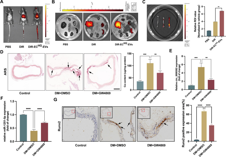
Fig. 5.
ECHG-EVs mediated arterial calcification in diabetic mice. A–C Fluorescence signals were detected in the living mice and organs (aorta, heart, lung, kidney, liver and spleen), respectively, after 12 h post-injection of DiR-labeled ECHG-EVs in mice. D ARS staining analysis showed the arterial calcification in different mice (n = 5/group). The arrows indicate calcified arteries (scale bar = 200 μm). E–F The expression of circ_0008362 and miR-1251-5p in different mice arterial tissues (n = 5/group). G Immunohistochemistry staining analysis of Runx2 expression in different mice arterial tissues. The arrows indicate the positive expression of Runx2 in the mice aorta (scale bar = 50 μm). One-way ANOVA with Tukey’s multiple comparisons test (C–G) was used. Three independent experiments were performed, and data were shown as mean ± SD. ****p < 0.0001, ***p < 0.001, **p < 0.005. ECHG-EVs: EVs derived from high-glucose induced ECs; DM: diabetes mellitus; DMSO: Dimethyl sulfoxide; ARS: Alizarin red s

