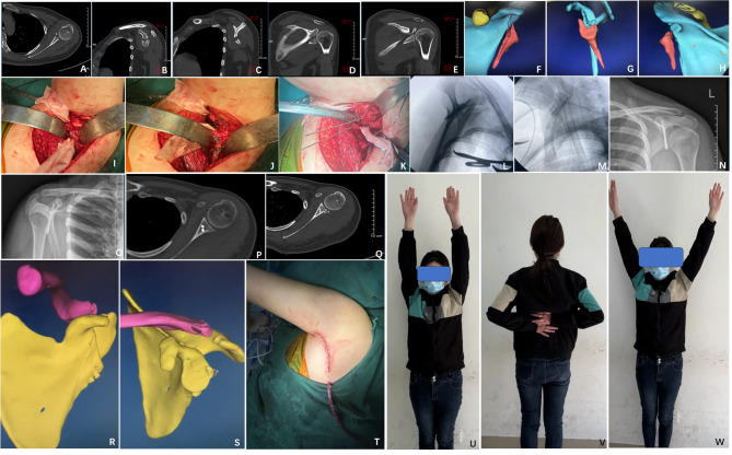
Fig. 3.
A 32-year-old female patient with left scapular glenoid fracture due to a vehicle accident, classified as Ideberg Type II. A ∼ H Preoperative CT scan showed scapular glenoid fracture(L)with fracture extending to the distal region; I The proximal fragment of the fracture was fixed with screws during surgery; J The distal fragment of the fracture was fixed using a Nice-Knot technique; K Surgical exposure through the axillary approach; L ∼ M intraoperative fluoroscopy showed excellent fracture reduction and proper screw placement; N ∼ S postoperative X-ray and CT scan showed excellent fracture reduction, satisfactory screw fixation without entering the joint cavity; T appearance of the incision site through the axillary approach, concealed and aesthetically pleasing; U ∼ W The patient’s shoulder joint function recovered well at the 6-month follow-up after surgery

