. 2024 Oct 18;105(10):002032. doi: 10.1099/jgv.0.002032

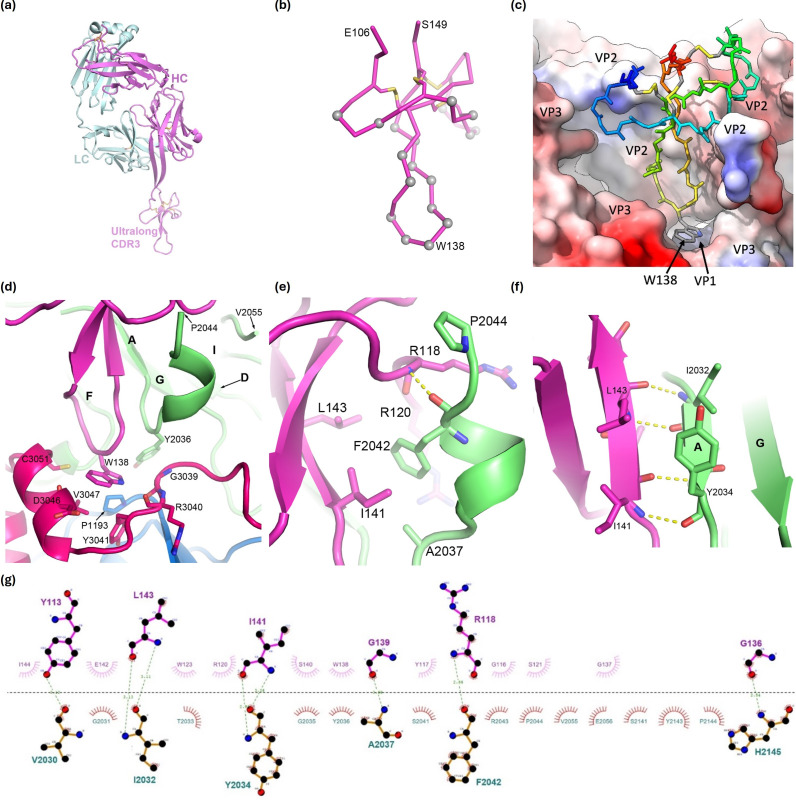
Fig. 3. Details of Fab117 binding. For clarity, only interacting residues and bonds are shown. Residues are labelled with those from the virus given a preceding chain identifier for VP1, VP2 or VP3, respectively, such that they all have four digits. (a) AlphaFold model of Fab117 with HC in magenta and LC in cyan. (b) Cα trace of the ordered ‘knob’ domain of CDR-H3 in the SAT2 pentamer–Fab117 complex. Cαs of residues contacting the antigen (within 4.0 Å) are shown as grey spheres and disulphides as yellow sticks. The CDR-H3 is orientated as in (a). (c) Fab117 (rainbow backbone) engages the hydrophobic cleft of the SAT2 pentamer shown in surface representation and coloured by hydrophobicity. (d) The tip of the Fab117 CDR-H3 is shown in purple with W138 central and SAT2 residues within 4.0 Å of the latter represented as sticks. VP2 is coloured green, VP3 red and the antibody purple, with VP2 secondary structural elements labelled. (e, f) Zoomed perspective of the interactions between Fab117 and VP2. VP2 secondary structural elements are labelled. (g) LigPlot+v2.2.8 [38] of the interaction between the Fab117 CDR-H3 and interface residues of VP2 only (thus, for instance, the contacts of W138 to VP1 and VP3 are not shown).

Fig. 3.