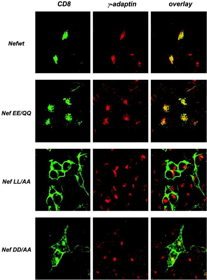
FIG. 3.
Subcellular distribution of the CD8-Nef chimeras and AP1 complexes. HeLa cells were transfected with the CD8-Nef expression vectors. Twenty-four hours later, cells were fixed and permeabilized. The CD8-Nef chimeras were stained with fluorescein isothiocyanate-conjugated anti-CD8 MAb (green), and AP1 was detected with anti-γ-adaptin followed by staining with a Cy3-conjugated Fab fragment recognizing mouse immunoglobulin G (red). Images were collected using a 63× oil immersion objective.

