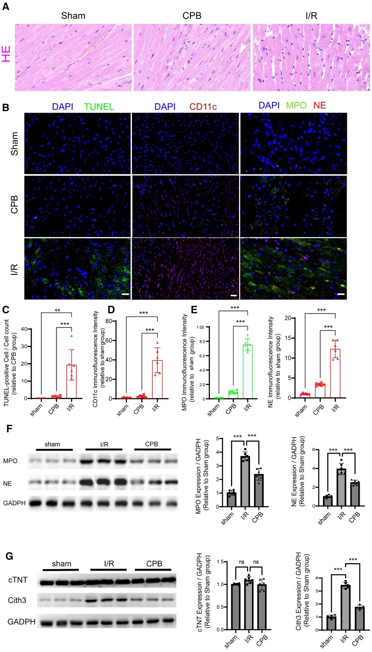
Figure 3:
NETs release increased in the perioperative period of heart surgery. (A) HE staining assessed inflammatory cell infiltration in myocardial tissues from sham, CPB and I/R rats. (B) TUNEL staining (scale bars, 20 µm) and immunofluorescence for CD11c, MPO and NE (scale bars, 20 µm) evaluated apoptosis and protein expression levels. (C) Apoptosis is represented as the ratio of TUNEL-positive cells to total cells (n = 6). (D and E) Fluorescence intensity of CD11c, MPO and NE was semi-quantitatively analysed (n = 6). (F and G) Western blot quantified MPO, NE, cTNT and CitH3 levels in myocardial tissues (n = 6). ‘ns’ denotes no significant difference. **P < 0.01, ***P < 0.001. Data are expressed as mean (SD). CitH3: citrullinated histone H3; CPB: cardiopulmonary bypass; cTNT: cardiac troponin T; HE: haematoxylin and eosin; I/R: ischaemia–reperfusion; MPO: myeloperoxidase; NE: neutrophil elastase; NETs: neutrophil extracellular traps; SD: standard deviation.

