Fig. 9.
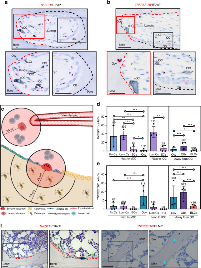
TNFSF11 and TNFRSF11B expression in human cortical and trabecular bone. a–e Data is obtained from cortical femoral bone specimens acquired from 4 adolescent females, and (f) from iliac crest bone sections from healthy humans. a Representative image of a selected cutting cone from TNFSF11 ISH (red), combined with a TRAcP (black) immunostained section. b Representative images of a selected cutting cone from TNFRSF11B (red) ISH, combined with a TRAcP (black) immunostained section. Red and black dashed lines in (a, b), respectively indicate eroded and formative surfaces, as determined by Masson’s Trichrome stained adjacent sections. c Methodological approach. d Histograms illustrate the mean percentage of TNFSF11+ cells located either next to surface osteoclasts, away from surface osteoclasts, or next to lumen osteoclasts. e Histogram illustrating the mean percentage of TNFRSF11B+ located either next to surface osteoclasts, away from surface osteoclasts, or next to lumen osteoclasts. The number above each bar is the number of cells quantified. f Representative images of human iliac crest bone sections stained for TNFSF11 or TNFRSF11B ISH (red), combined with a TRAcP immunostaining (black). TRAcP+ Osteoclasts (black arrow), TNFSF11+ Canopy cells (red arrows), reversal cells (green arrows), marrow cells (purple arrows), and TNFRSF11B+ osteocytes (blue arrows). d, e Data are shown as mean ± SD. Clustered logistic regression was utilized to determine the likelihood of TNFSF11 or TNFRSF11B being expressed in one cell population compared with another. ***P values < 0.001, **P values < 0.01, *P values < 0.05. P values, odds ratios, and corresponding 95% confidence intervals are shown in Table S1. Scale bars: overview = 100 µm, high magnification images = 50 µm. sOC surface osteoclast, lOC lumen osteoclast, Ocy osteocyte, OB osteoblast, Rv.C reversal cell, Lum.C lumen cell, EC vascular endothelial cell, BLC bone lining cell, Illustrations were created using BioRender.com

