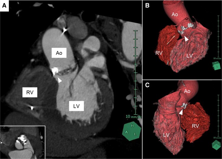
Figure 2.
Calcified aortic valve complex on computed tomography. (A) Left anterior oblique cross-sectional image reveals heavy aortic valve calcification extending to the interventricular membranous septum. Inset image shows a view parallel to the aortic valve. (B and C) 3D images reconstructed from computed tomography. White arrowheads indicate calcification attached to aortic valve. Ao, ascending aorta; LV, left ventricle; RV, right ventricle.

