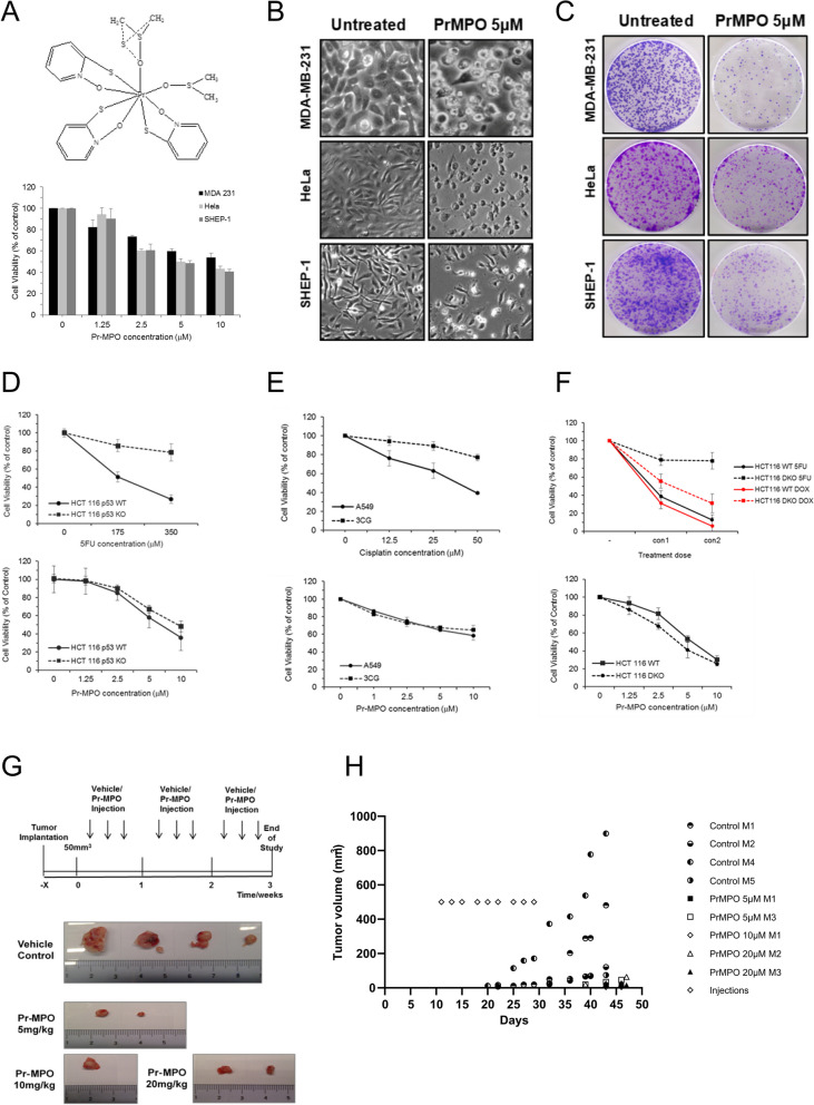
Fig. 1.
Pr-MPO induces cell death in a wide range of cancer cell lines. A The top panel shows the structure of Pr-MPO (praseodymium (2-mercaptopyridine N-oxide) (dimethyl sulfoxide)). The lower panel shows the effect of a dose response of Pr-MPO on HeLa, MDA-MB-231 and SHEP-1 cells assessed by crystal violet staining. B Light microscope observation and C Colony forming assay of HeLa, MDA-MB-231 and SHEP-1 cells untreated or treated with Pr-MPO 5µM. Comparison of cell sensitivity assessed through crystal violet staining D of HCT116 p53 wild type or p53 KO to increasing concentrations of 5FU or Pr-MPO, E of HCT116 wild type and HCT1116 Bax−/− Bak−/− to increasing concentrations of 5FU, doxorubicin and Pr-MPO and F of A549 and 3CG to increasing concentrations of cisplatin and Pr-MPO. G Xenograft protocol used to assess the ability of DU145 cells to form tumors in nude mice (top panel) and pictures of tumors collected from control and Pr-MPO treated animals (lower panel). H Evolution of tumor volumes in control and Pr-MPO treated animals

