
FIGURE 2.
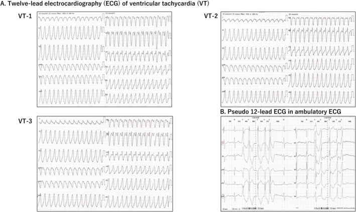
(A) Twelve‐lead electrocardiography (ECG), (B) Pseudo‐12‐lead ECG in ambulatory ECG. Sustained ventricular tachycardia (VT)‐1, VT‐2, and VT‐3 shows 176–190 bpm, left bundle branch block, inferior axis, and transition zone of V3.
Official websites use .gov
A
.gov website belongs to an official
government organization in the United States.
Secure .gov websites use HTTPS
A lock (
) or https:// means you've safely
connected to the .gov website. Share sensitive
information only on official, secure websites.
(A) Twelve‐lead electrocardiography (ECG), (B) Pseudo‐12‐lead ECG in ambulatory ECG. Sustained ventricular tachycardia (VT)‐1, VT‐2, and VT‐3 shows 176–190 bpm, left bundle branch block, inferior axis, and transition zone of V3.