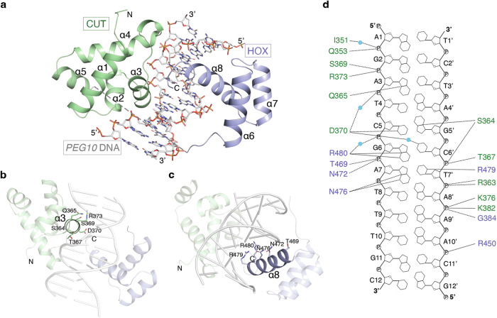
Fig. 1. Structure of the OC2-PEG10 complex.
a Overall structure of the OC2-PEG10 DNA complex. The position of CUT and HOX domains on DNA and their respective helices are labeled (α1- α8) while the unmodelled loop is depicted as a dashed line. CUT and HOX domains are shown in green and blue, respectively. b, c Arrangement of DNA interacting residues in α3 helix of CUT domain and α8 helix of HOX domain of OC2 are shown. d Schematic representation of the protein-DNA contacts in the complex. Hydrogen bonds are shown as dashed lines and water molecules are depicted as cyan spheres. DNA interacting residues of CUT and HOX domains are shown in green and blue, respectively.

