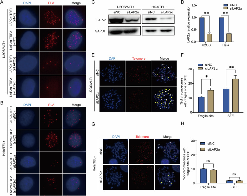
Fig. 1. LAP2α interacted with shelterin complex and LAP2α deficiency induced dysfunctional telomeres of ALT-positive cells.
A, B PLA of LAP2α and subunit of shelterin complex in U2OS cells and Hela cells transfected with control or LAP2α siRNA. Red dots represent PLA signals. C U2OS and Hela cells were infected with control or LAP2α siRNA for 3 days. Cell lysates were subjected to western blot analysis with anti-LAP2α and anti-GAPDH antibodies. GAPDH was used as the loading control. D Quantification of the LAP2α relative protein level in comparison to GAPDH and to siNC was calculated by ImageJ software. Error bars represent the mean ± SEM of four independent experiments. Two-tailed unpaired Student’s t-test was used to calculate p-values. **p < 0.01. E, G Metaphase chromosome spreads and telomere FISH in U2OS and Hela cells. Representative images showing fragile sites (yellow arrows) and signal-free ends (SFE, white arrows). F, H Quantification of the fragile site and SFEs of U2OS and Hela cells. Error bars represent the mean ± SEM of three independent experiments. Two-tailed unpaired Student’s t-test was used to calculate p-values. ns, not significant or p ≥ 0.05; *p < 0.05; **p < 0.01.

