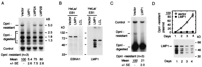
FIG. 1.
Expression of LMP1 suppressed replication of the oriP plasmid in HeLa/EB1 cells. (A) Transient replication assay of the oriP plasmid. The oriP plasmid (2 μg), the control plasmid (1 μg), and the expression plasmid of LMP1, LMP2A, or LMP2B (0.5 μg) were transfected. Total amounts of plasmids were adjusted to 3.5 μg with the vector plasmid. Hirt's extracts were prepared 2 days after transfection, and plasmids were analyzed by DpnI digestion and Southern blot hybridization. The linearized DpnI-resistant plasmid (5.0 kb) and three DpnI-digested fragments (2.5, 1.8, and 1.3 kb) are shown. Two fragments indicated by asterisks (2.5 and 1.3 kb) are products of replication intermediates that were accumulated by the replication fork barrier at the FR element of oriP. Amounts of the DpnI-resistant plasmid (replicated plasmid) are normalized with that of the control plasmid and shown below. Data represent averages of three experiments with the standard errors (SE). (B) Expression of LMP1 and EBNA1 in transfected cells. Short polypeptides reacted with LMP1 antibody were digested products of LMP1. As a control, a similar number of LCL cells were analyzed in a parallel lane. (C) Replication assay of the DS plasmid. Experimental conditions are described above for panel A. Data represent averages of three experiments with the standard errors (SE). (D) Time course of the oriP plasmid replication. Transfected plasmids were the same as in panel A, and cells were collected for DpnI assay at the days indicated. A summary of two experiments is shown. Expression of LMP1 in one experiment is shown at the bottom. The same amount of total proteins was loaded on each lane.

