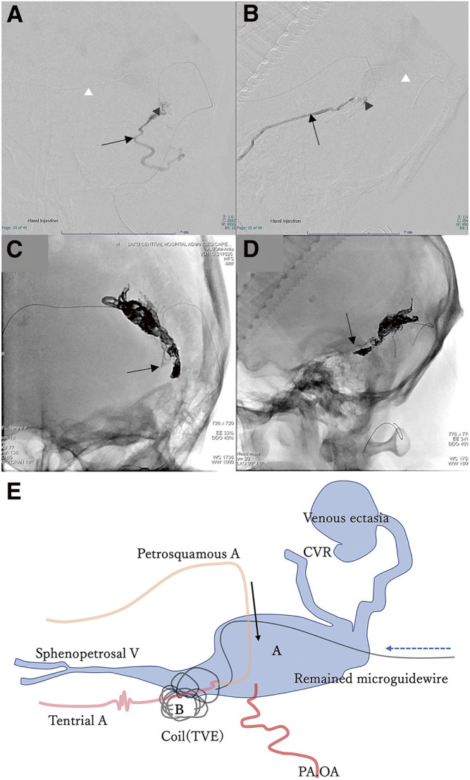
Fig. 4. (A–D) Intraoperative DSA via microcatheter during TAE, frontal view (A and C), and lateral view (B and D). Microcatheter is inserted via the petrosquamous branch of the middle meningeal artery into the left transverse sinus through the shunt point (A and B, black arrowhead). Sphenopetrosal vein (A and B, black arrow). Microguidewire remnant after fracture (A and B, white arrowhead). Onyx cast front end (C and D, black arrow). (E) Illustration of the treatment progress. A: left transverse sinus, B: left sigmoid sinus, black arrow: approach direction during TVE, dotted line arrow: approach direction during TVE. CVR, cortical venous reflux; OA, occipital artery; PA, posterior auricular artery; TAE, transarterial embolization; TVE, transvenous embolization.

