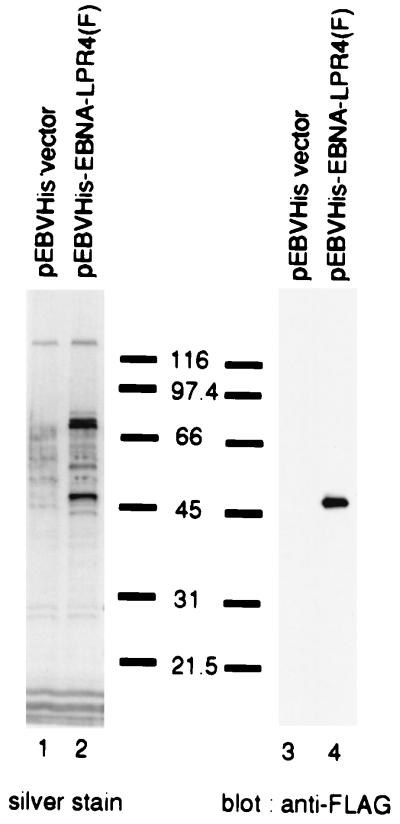
FIG. 3.
Photographic images of a silver-stained denaturing gel (left panel) and an immunoblot (right panel) of electrophoretically separated cell proteins bound to anti-FLAG monoclonal antibody (M2)-conjugated agarose beads. BOSC23 cells were transiently transfected with the empty vector (lanes 1 and 3) or pEBVHis-EBNA-LPR4(F) (lanes 2 or 4), harvested, solubilized, and reacted with anti-FLAG monoclonal antibody (M2)-conjugated agarose beads. The beads were pelleted, rinsed extensively, and eluted with the FLAG peptide. The eluted fractions were then electrophoretically separated in duplicate on two denaturing gels. Each gel was subjected to silver staining (left panel) or immunoblotting with anti-FLAG monoclonal antibody (M2) (right panel). The band corresponding to EBNA-LP was cut out, digested with trypsin, and subjected to mass-spectrometric analysis as described in the text.

