FIG. 6.
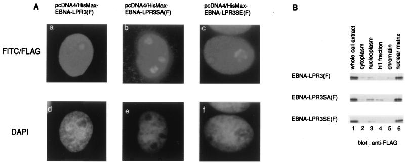
Subcellular localization of EBNA-LP mutants. (A) COS-7 cells were transiently transfected with the expression vectors described in Fig. 4A and subjected to an indirect immunofluorescence assay as described in Materials and Methods. The expressions of EBNA-LPs were visualized with anti-FLAG monoclonal antibody (M2) and fluorescein isothiocyanate conjugated anti-mouse IgG antibody (a to c). DNA was visualized with DAPI (d to f). (B) BOSC23 cells were transiently transfected with pME-EBNA-LPR3(F) (upper panel), pME-EBNA-LPR3SA(F) (middle panel), and pME-EBNA-LPR3SE(F) (lower panel) and fractionated as described in Materials and Methods. Each fraction (lane 1, whole extract; lane 2, cytoplasmic fraction; lane 3, nucleoplasmic fraction; lane 4, H1 fraction; lane 5, chromatin fraction; lane 6, nuclear matrix fraction) was solubilized, separated in denaturing gels, and subjected to immunoblotting with anti-FLAG monoclonal antibody (M2).

