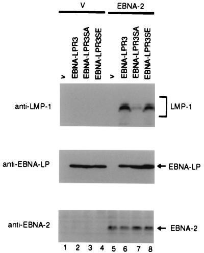
FIG. 7.
Photographic images of immunoblots of electrophoretically separated Akata cells transiently transfected with various EBNA-LP expression vectors in combination with pME18S (V) (lanes 1 to 4) or pME-EBNA-2 (EBNA-2) (lanes 5 to 8). At 48 h posttransfection, cells were harvested, solubilized, and subjected to immunoblotting with anti-LMP1 monoclonal antibody (S-12) (upper panel), anti-EBNA-LP monoclonal antibody (JF186) (middle panel), or anti-EBNA-2 monoclonal antibody (PE2) (lower panel).

