
Figure 3. In Silico modeling predicts enhanced T cell priming with extended-prime immunization, consistent with experimental measurements of DC antigen acquisition and activation in draining lymph nodes.
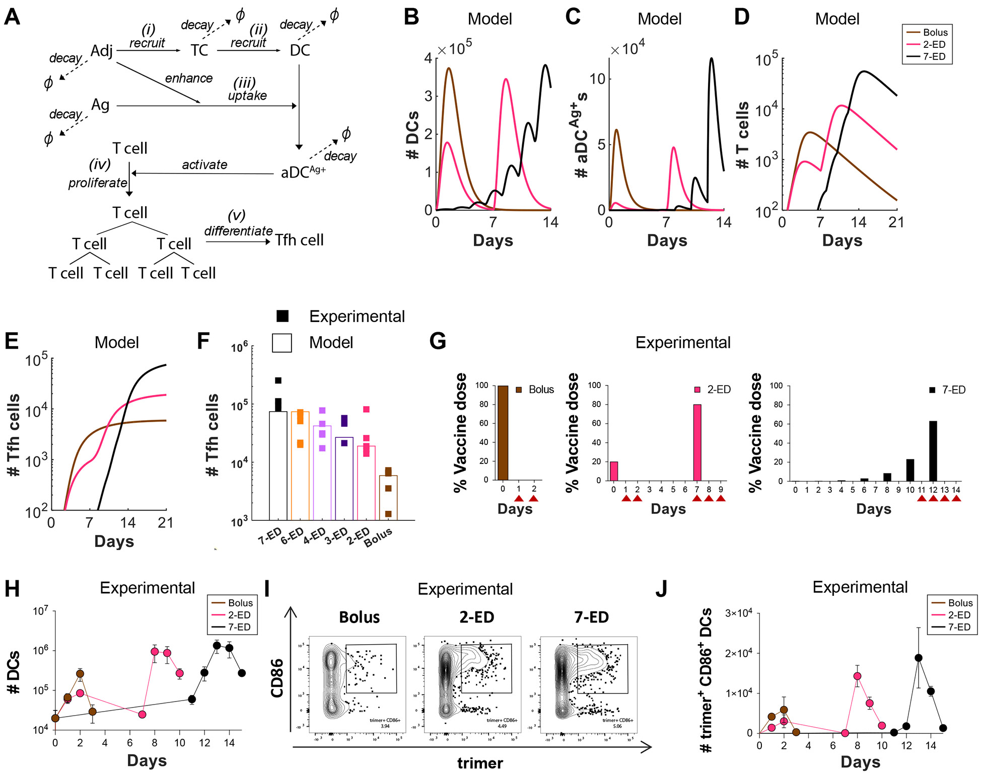
(A-F) Computational model of vaccine uptake by dendritic cells and helper T cell priming. (A) Schematic outlining elements of the kinetic model. (B-E) Modeling predictions of the number of (B) total DCs (B), Ag+Adj+ DCs (C), Ag-specific T cells (D), and Tfh cells (E) for bolus, 2-ED, or 7-ED immunization regimens. (F) Comparing Tfh cell count predicted by the model with the experimental data at day 14. (G-J) Experimental analysis of lymph node DC antigen uptake and activation. C57BL/6J mice (n=3 animals/group) were immunized with 10 μg Cy5 dye-labled-N332-GT2 trimer and 5 μg SMNP adjuvant according to the dosing schemes shown in (G), and DCs in draining lymph nodes were analyzed by flow cytometry on days indicated by arrows. Shown are number of DCs (H), representative histograms of trimer antigen fluorescence and CD86 expression by CD11c+ DCs (I), and number of trimer+CD86+ DC counts over time for bolus, 2-ED, and 7-ED immunization regimens (J).