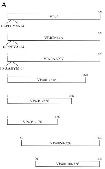
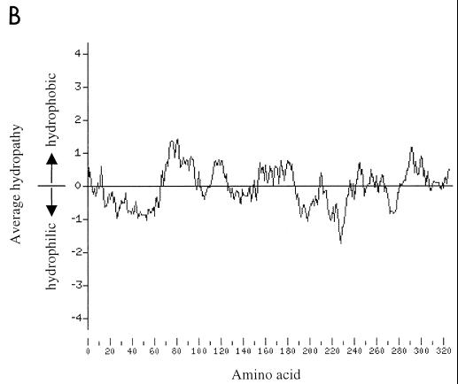
FIG. 1.
(A) Schematic representation of wild-type VP40 and VP40 mutant proteins reported in this paper. Amino acid residues are represented with the single-letter code. Substituted residues are indicated in boldface type. (B) Kyte-Doolittle hydrophobicity plot of Ebola virus VP40 over a window of 17 amino acids (16).


