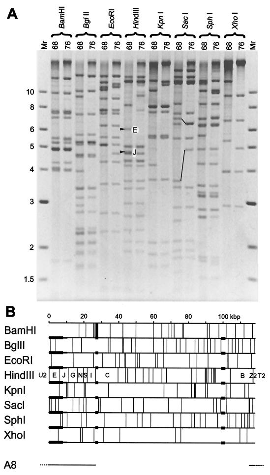
FIG. 1.
Structure of the MHV-76 genome. (A) Restriction endonuclease profiles of MHV-68 and MHV-76 virion DNAs. Samples were analyzed on a 0.8% agarose gel. Fragments E and J are indicated in the HindIII profile of MHV-68. These are absent from the HindIII profile of MHV-76. Fragments containing the 40-bp repeat (upper pair) and the 100-bp repeat (lower pair) are connected by lines in the SacI profiles of MHV-68 and MHV-76. Molecular size markers and their sizes in kilobase pairs are indicated to the left. (B) Restriction endonuclease maps of the unique region of the MHV-68 genome, highlighting the three regions that differ in MHV-76. Variable copy numbers of the terminal repeat (not shown) flank the unique region, resulting in heterogeneous populations of fragments from the genome termini differing by increments of 1.2 kbp. None of the restriction endonucleases for which maps are shown cleave the terminal repeat. Three regions in which MHV-76 and MHV-68 differ are denoted by thicker horizontal lines. Two correspond to variations in the copy number of reiterations in the 40-bp repeat (at kbp 27) and 100-bp repeat (at kbp 100). The third represents a deletion at the left end. Minimal and maximal extents of the deletion as deduced from panel A are indicated by thicker and thinner lines, respectively. The location of the insert in MHV-68 cosmid A8 is indicated at the bottom, with terminal repeat sequences dashed, and the corresponding fragment nomenclature is included in the HindIII map.

