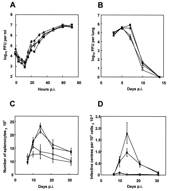
FIG. 7.
Biological characterization of MHV-68, MHV-76, and the rescuant viruses. Data points (mean value ± standard error) are shown in each graph for MHV-68 (▪), MHV-76 (○), MHV-76(cA8+)4 (▴), and MHV-76(cA8-)5 (▵). (A) Single-step growth curves comparing growth of viruses on BHK-21 cells at an MOI of 5. The data are representative of two separate experiments, and each experiment was done in duplicate. (B) Viral replication in the lungs of BALB/c mice infected intranasally with 2 × 10⁵ PFU of virus. Data for four mice per group are shown at each time point. (C) Numbers of spleen cells during intranasal infection of BALB/c mice with 2 × 10⁵ PFU of virus. Data for four mice per group are shown at each time point. (D) Latent virus in the spleens of BALB/c mice infected with 2 × 10⁵ PFU of virus as determined by infective-center assay. Infectious virus titers (less than 50 PFU per spleen in every case) have been subtracted from the infective-center results. Data for four mice per group are shown at each time point.

