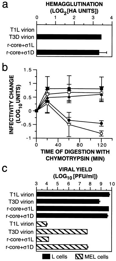
FIG. 5.
Capacity of r-cores+ς1 to mediate ς1-specific in vitro and in vivo properties. (a) HA of bovine RBCs (Colorado Serum Co.) induced by purified T1L virions, T3D virions, r-cores+ς1L, and r-cores+ς1D was determined as described for Fig. 3a. Averages ± standard deviations of three replicates are shown. (b) The effect of chymotrypsin treatment on infectivity of T1L virions (open squares), T3D virions (open circles), r-cores+ς1L (filled squares), r-cores+ς1D (filled circles), and r-cores+ς1D(T249I) (filled triangles) was determined as follows. Purified virus (1.5 × 10¹² particles/ml) was incubated with chymotrypsin (200 μg/ml) at 37°C. At specified times, an aliquot was removed onto ice and digestion was stopped with phenylmethylsulfonyl fluoride (2 mM). The infectivity of each aliquot was determined by plaque assay. Viral infectivity at a specified time (t) relative to time zero was expressed as described for Fig. 4c. Averages ± standard deviations of three independent experiments are shown. (c) Infectious yields produced in MEL and L cells by T1L virions, T3D virions, r-cores+ς1L, and r-cores+ς1D were determined as follows. Purified virus (5 PFU/cell) was incubated with 4 × 10⁶ cells for 1 h at room temperature. Unbound virus was removed by centrifugation (500 × g) and washing cells with phosphate-buffered saline. Cells were then resuspended in growth medium and incubated at 37°C for 24 h. Cell lysates were generated by freeze-thawing, and the amount of infectious virus in these lysates was determined by plaque assay. Growth yields were expressed as log10(PFU/ml) at 24 h. Averages ± standard deviations of six trials from two independent experiments are shown.

