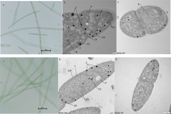
Fig. 1.
Morphology and ultrastructure of Aphanizomenon sp. KUCC C1 and KUCC C2 cells: light microscopy of KUCC C1 (A) and KUCC C2 (D), ultrathin TEM sections: general view of the longitudinal section of KUCC C1 (B) and KUCC C2 (E) cells, and dividing cells of KUCC C1 (C) and KUCC C2 (F). Cyanophycin granules (C), polyphosphate granules (P), phycobilisomes (PH), cell wall (CW), cross wall (CR), nucleoplasm (N), intrathylakoid space (ITS), thylakoids (T), carboxysome (CS). Scale bars: 25 μm (A, D), 200 nm (B, C), 500 nm (E), 1 μm (F).

