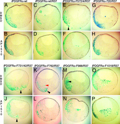
Fig. 2.
PDGFR signaling through PLCγ and PI3K, but not through SHP-2, Shf, and Crk, is required for mesoderm cell survival. (A and B) Embryos were coinjected with mRNA encoding β-gal and iPDGFRα or β-gal, PDGFR-37 (R37), and the following iPDGFRαs. (C and D) wt. (E and F) F572/74. (G and H) F720. (I and J) F731/42. (K and L) F762. (M and N) F988. (O and P) F1018. At the beginning of gastrulation (stage 10), AP1510 or DMSO was injected into the blastocoel. At the midgastrula stage (stage 11), β-gal expression was visualized (shown in blue). The stained embryos were dissected and scored for the presence or absence of nonnuclear β-gal-stained cells in the blastocoel cavity, within the vitelline membrane, or in the process of being excluded from the embryo (see red arrowhead in K), indicating the presence of apoptotic cells. The percentage of embryos containing apoptotic cells was calculated. Representative saggital sections of these embryos are shown. (G–P) Note that when signaling through PLCγ and PI3K is prevented (I, J, and M–P), activation of the receptor with AP1510 did not restore cell survival, whereas cell survival is restored when signaling through SHP-2, Shf, and Crk is prevented (G, H, K, and L). Arrowheads indicate apoptotic mesoderm cells outside the blastocoel cavity. Note that overexpression of iPDGFRα-wt mRNA alone does not cause apoptosis in mesoderm cells.

