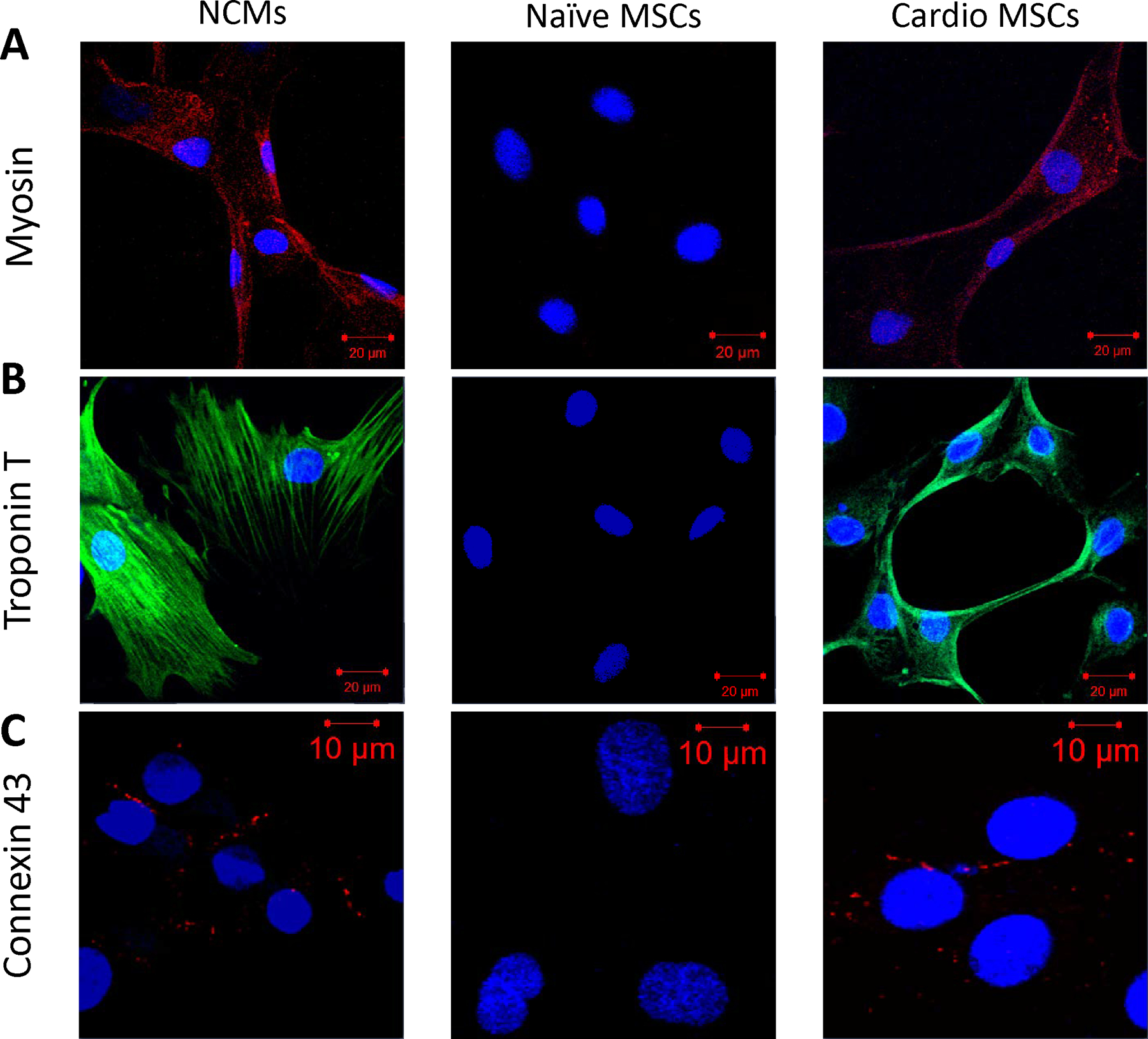
Figure 7.

Immunofluorescence staining for cardiac proteins. NCM (left), naïve MSCs (middle) and cardio MSCs (right column) were stained for (A) MHC (Texas Red), (B) TropT (FITC) and (C) Cx43 (Texas Red) and counterstained with DAPI. Cardio MSCs showed increased expression of cardiac proteins, indicating a shift to a cell with cardiomyogenic characteristics. Slides were photographed on a laser scanning confocal microscope at 20x magnification.
