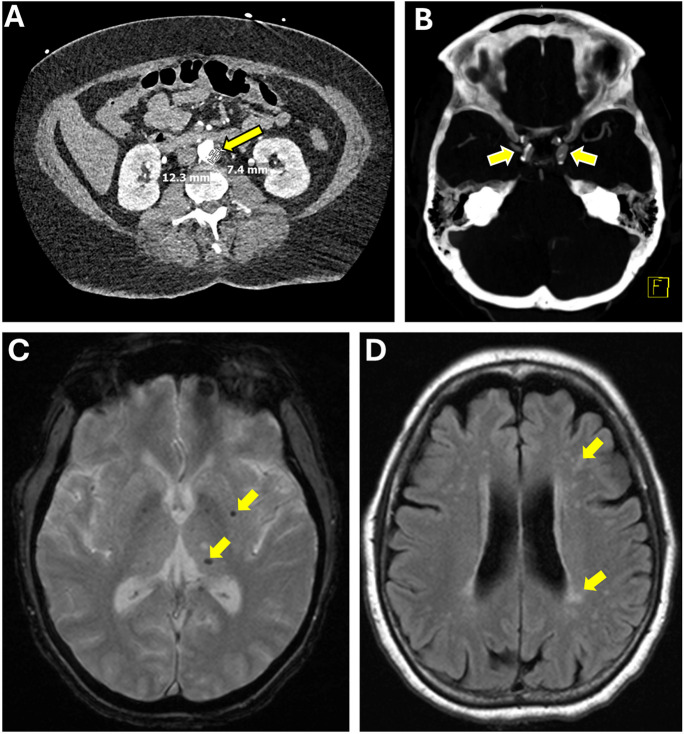
Fig. 4.
The intersection of age-related peripheral and cerebral vascular disease: a case study. This figure displays the medical journey of a 67-year-old female with a history of hypertension, non-insulin-dependent diabetes mellitus, and significant infrarenal aortic atherosclerosis (A, arrow) leading to severe stenosis of the left femoral artery, who experienced dizziness and nausea. Imaging revealed an acute right cerebellar infarct and right vertebral artery occlusion, suggestive of acute atherosclerosis or dissection. CT angiography further identified bilateral intracranial carotid artery stenosis due to atherosclerosis (B), while MRI GRE sequences showed microhemorrhages (C, arrowheads), and FLAIR sequences highlighted white matter hyperintensities (D), indicative of chronic small vessel disease. This case encapsulates the intricate connection between systemic atherosclerotic disease and its cerebral manifestations, demonstrating how atherosclerosis can precipitate both acute cerebrovascular events and chronic small vessel disease

