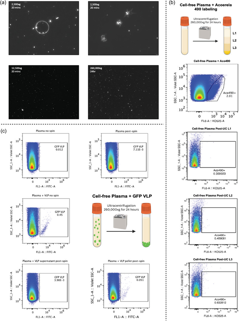
FIGURE 2.

Extracellular vesicles are largely found in the pellet as opposed to supernatant following ultracentrifugation. Cell‐free plasma was processed through ultracentrifugation 260,000 × g overnight and analysed through dark‐field microscopy and flow cytometry with fluorescence labelling. (a) Dark field microscopy also revealed marginal differences in plasma supernatant content, due to large abundance of lipoproteins, with the following centrifugation parameters: 2500 × g for 20 min, 12,500 × g for 20 min, or 260,000 × g for 24 h. (b) Following centrifugation, use of EV‐specific membrane fluorogenic dye – Acoerela 490 – revealed almost no presence of EVs in the supernatant, which was aliquoted into three separate layers from top to bottom. For flow cytometry analysis, a total of 25,000 events were acquired in EV gate under Violet SSC trigger. (c) To confirm this finding, standard GFP‐labelled viral‐like particles (VLPs) were spiked into cell‐free plasma prior to centrifugation. Both plasma with and without GFP VLPs were analysed, either stored overnight in 4°C or following ultracentrifugation at 4°C. A total of 250,000 events were acquired from EV gate under Violet SSC trigger. Comparison between the supernatant and pelleted particles following ultracentrifugation reveals that most EVs are located within the pellet as opposed to the supernatant.
