FIGURE 3.

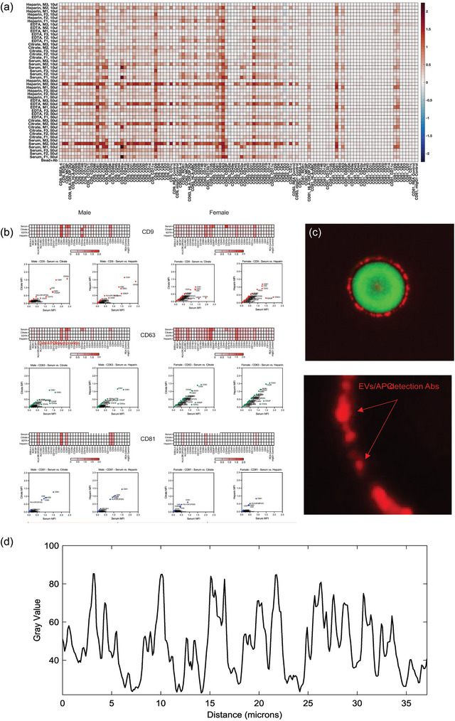
Multiplex assay reveals relative similarities in surface epitope profiles of EVs from different anticoagulants (ACS). Surface antigen profiling of EVs in cell‐free plasma collected with the four different ACs was performed with the MACSPlex Human Exosome Multiplex Assay kit, consisting of 39 unique fluorescently barcoded capture beads and three detection antibodies (CD9, CD63, CD81). Cell‐free plasma was collected from three female and three male donors, with preparation of two sample volumes for analysis (50 and 10 µL). MPAPASS was utilized for data analysis, revealing more inter‐donor differences as opposed to inter‐AC differences. (a) A heat map was generated, showing the expression levels of antigens for each sample from low (blue) to high (dark red), for three female and three male donors. (b) Correlation plot analysis was also performed to demonstrate the correlations between ACs that were largely similar based on the heat map, such as heparin and serum. (c) The signal originating from captured EVs (red fluorescence) does not allow for identification of EVs positive for the capture Ab target, or specific analysis of EV size and their respective antigenic expression levels (arrow in c). (d) Relative intensity histogram, showing significant variation of the CD9 signal across the captured EVs. Bar represents 3 and 1 um, respectively.
