
Figure 1.
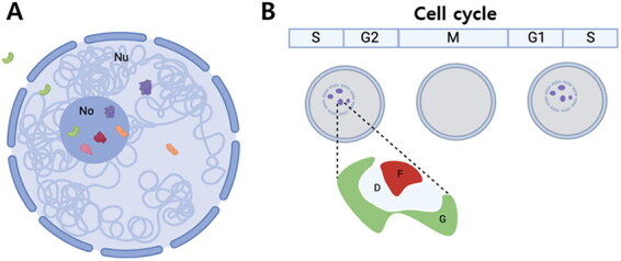
Schematic diagram showing dynamics of nucleolar proteins (A) and cell cycle-dependent changes in nucleoli (B). Some of the nucleolar proteins (red and pink) reside, functioning mainly in nucleolus, and others move between nucleoplasm and nucleolus (purple and orange) (a). They shuttle between nucleoplasm and nucleolus, either because they should be sequestered inside the nucleolus until they are required by the nucleus, or because they have major roles in the nucleolus and have moonlighting functions in the nucleoplasm. There are even proteins that go out to the cytoplasm (green). Nucleoli undergo disassembly and assembly during the cell cycle (B). The figures are created based on the mammalian nucleoli, which have tri-partite structure consisting of fibrillar center (F), dense fibrillar center (D), and granular component (G).