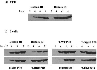
FIG. 3.
Western blot analysis of expression of M1 in influenza virus-infected CEF (a) and L (b) cells. CEF cells or L cells were infected at high MOI with Dobson 4H, Rostock S3, or transfectant viruses T-WT PB2, T-tagged PB2, T-RD1 PB2, T-RDR, T-RDR1968, or T-RDR2128. At the time points indicated, cells were washed once in ice-cold PBS and incubated with urea-SDS lysis buffer on ice for 10 min. Cell lysates were separated by SDS–15% PAGE and probed for M1 with mouse anti-influenza A virus matrix protein and anti-mouse horseradish peroxidase-conjugated secondary antibodies. Proteins were visualized using an ECL detection system.

