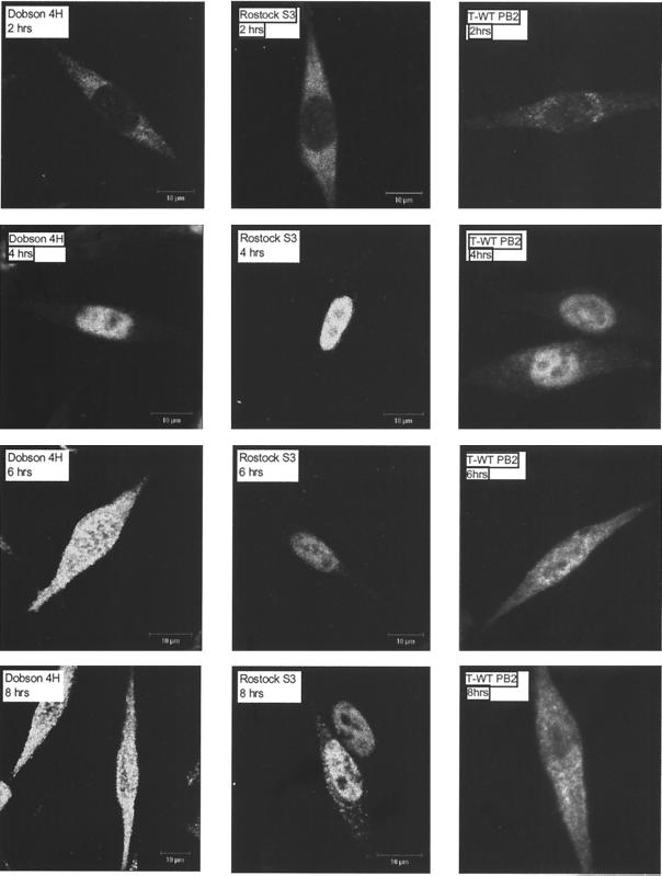
FIG. 4.
Localization of NP in L cells during infection. L cells were grown on glass coverslips to 50 to 70% confluency and infected at high MOI with Dobson 4H, Rostock S3, or T-WT PB2. At 2, 4, 6, and 8 h postinfection, cells were processed for immunofluorescence analysis by fixation with 4% paraformaldehyde in PBS for 10 min and then permeablized with 0.2% Triton X-100 in PBS for 5 min. Cells were stained with a mouse anti-influenza A virus nucleoprotein monoclonal antibody and an anti-mouse immunoglobulin G-fluorescein isothiocyanate conjugate. Fluorescence was viewed with a confocal microscope.

