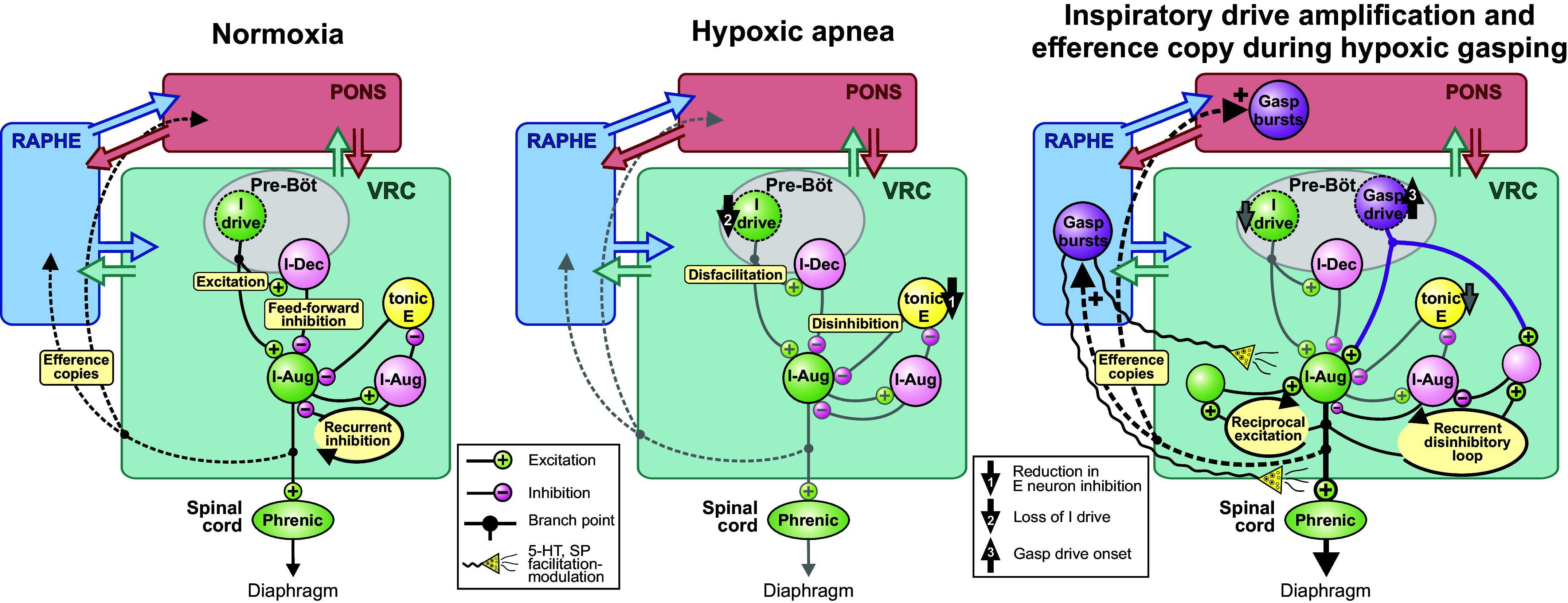
Figure 6.

Graphical summary of hypotheses and circuit operations suggested by the results. Left: simplified model of functional correlations and circuit mechanisms during normoxia. E, expiratory; I, inspiratory; pre-Böt, pre-Bötzinger complex; VRC, ventral respiratory column. Center: a reduction in expiratory neuron inhibition and a loss of inspiratory drive (arrows 1 and 2), respectively, result in augmentation and depression of inspiratory drive, leading to hypoxic apnea. Right: the onset of gasp-driver neuron bursting (arrow 3) excites downstream inspiratory premotor neurons. Efference copies (i.e., corollary discharge such as that which occurs with axon collaterals) of the inspiratory drive engage raphe and pontine circuits in a distributed network of coordinated gasp-related bursting (right; dashed lines) in an attempt to generate autoresuscitative efforts. See text for further discussion. I-Dec (I-Aug), peak neuronal firing rate occurred during the first (second) half of the inspiratory phase; SP, substance P.
