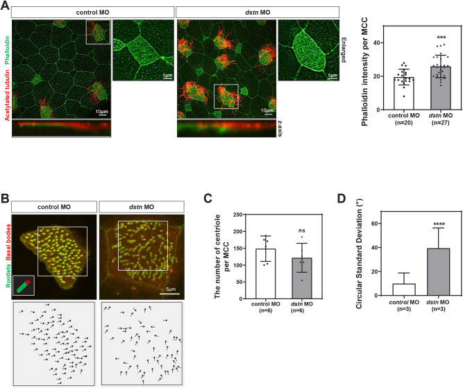
Fig. 2. Dstn is required for ciliogenesis during Xenopus embryonic development.
(A) Xenopus epidermal MCCs marked by immunofluorescence using anti-acetylated tubulin (red) for cilia and phalloidin (green) for actin filament network (scale bar=10 μm; enlarged scale bar=5 μm). Statistical quantitation of phalloidin intensity revealed a significant increase in phalloidin intensity per MCC in dstn-depleted embryos as compared to control embryos. (B) Centrin-RFP and Clamp-GFP exhibited perturbation of basal body polarity in dstn-depleted embryos compared with coordinated polarity and alignment in control embryos. It is highlighted by black arrows (scale bar=5 μm). (C) We have quantified the number of centrioles per MCC in Xenopus epidermis and data showed that dstn knockdown did not significantly affect the number of centrioles in dstn morphant embryos. (D) The polarization was analyzed by angular measurements of Centrin/Clamp pairs in MCCs. Statistical analysis of data showed that dstn inhibition increased the circular standard deviation compared with control embryos (n=3). * p<0.05, ** p<0.01, *** p<0.001, **** p<0.0001, MO, morpholino oligonucleotides; MCC, multiciliated cells.

