
Fig. 2.
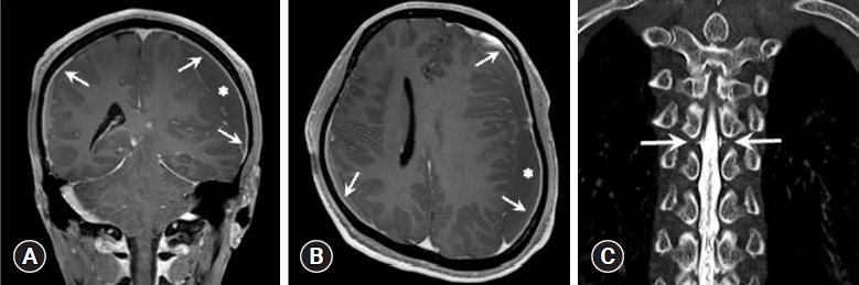
Case 2 images. (A, B) Gadolinium-enhanced T1-weighted magnetic resonance images of the brain showing diffuse pachymeningeal enhancement (arrows) and subdural left hemispheric chronic hematoma (asterisks). (A) Coronal view. (B) Axial view. (C) Computed tomography myelography coronal view showing leakage of contrast material (arrows) at T5 level.