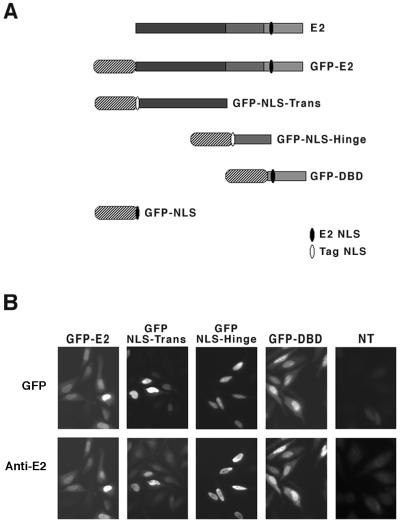
FIG. 1.
Schematic representation of the GFP fusion proteins and their expression in HeLa cells. (A) The three domains of E2 (gray bars) and the GFP moiety (stripped ovals) are shown. Sequence containing the E2 NLS is schematized in black, and the SV40 TAg NLS is shown in white. (B) The four constructs were transfected in HeLa cells, and their expression was checked 24 h later by either direct green fluorescence of GFP or immunofluorescence with anti-E2 antibodies revealed by Texas red as indicated. NT, not transfected.

