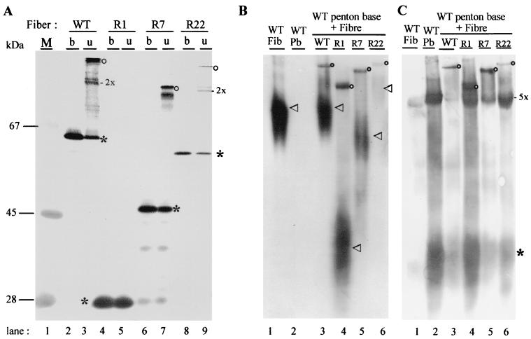
FIG. 2.
Recombinant fibers, ability to trimerize (A) and to form penton capsomers (B and C). (A) Fiber trimers were analyzed by SDS-PAGE and NDS-PAGE and immunoblotting with 4D2.5 antibody as boiled (b) or unboiled (u) samples. WT fiber (lanes 2 and 3), R1-RGD (lanes 4 and 5), and R7-RGD (lanes 6 and 7) were all highly expressed in Sf9 cells as soluble recombinant proteins, whereas R22-RGD was a low expresser (lanes 8 and 9). Note that no stable fiber trimer was detectable in unboiled R1-RGD lysate (lane 5). Fiber monomers are indicated by an asterisk, dimers by 2x, and trimers by an open circle. (B and C) Assembly of fiber with penton base was assayed in lysates of Sf9 cells coexpressing recombinant fiber and penton base (16). Native proteins were electrophoresed in an 8% nondenaturing gel and electrically transferred, and blots were reacted with antitrimer fiber 2A6.36 monoclonal antibody (B) or polyclonal anti-penton base (C). Control samples of single expression of WT fiber and penton base alone are shown in lanes 1 and 2, respectively. Lanes 3 to 6, lysates from cells coexpressing WT penton base with WT fiber (lane 3), R1-RGD (lane 4), R7-RGD (lane 5), or R22-RGD (lane 6), respectively. Penton capsomer, comprised of penton base-anchored fiber, migrates as a discrete band reacting with both antifiber 2A6.36 (B) and anti-penton base antibodies (C), and is indicated by open circles. In panel B, free fibers, migrating as a diffuse band of polydisperse species, are indicated by an arrowhead. In panel C, monomers of penton base are indicated by an asterisk and pentamers (penton base capsomers) by 5x.

