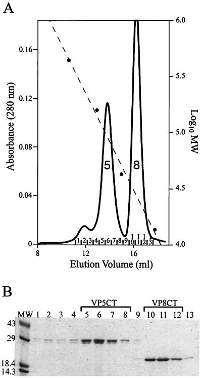
FIG. 4.
Purification of VP5CT and VP8CT. (A) Gel filtration chromatography. Shown is a chromatogram of VP5CT and VP8CT, produced by sequential digestion of VP4 with chymotrypsin and trypsin (protocol is described in Materials and Methods), separated on a Superdex 200 HR 10/30 column. The Vo is 8.43 ml. Solid line, A280; dashed line, calibration curve; circles, elution volumes of MW markers; peak 5, main VP5CT peak; peak 8, VP8CT peak. Fractions are numbered above the abscissa. (B) Coomassie blue-stained SDS-polyacrylamide gel. The fractions labeled in panel A were analyzed on a reducing 4-to-15% polyacrylamide gradient gel. Fraction numbers are indicated immediately above the lanes. Fractions pooled for further analysis of VP5CT and VP8CT are indicated above the brackets. Molecular mass standards are identified in kilodaltons adjacent to the marker bands.

