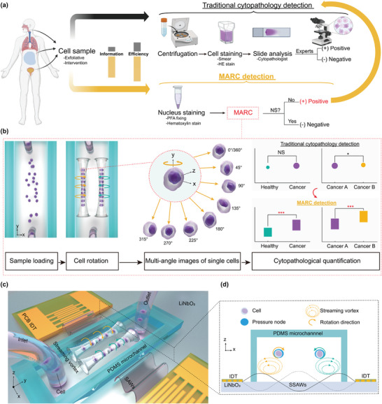
Figure 1.

Schematic of the multi‐view acoustofluidic rotation cytometry (MARC) for pre‐cytopathological screening. a) Cell samples collected from biofluid are subjected to two analysis pathways. The traditional path involves sample centrifugation, hematoxylin and eosin (HE) staining and cytopathologist examination, yielding binary outcomes based on experience. Alternatively, the MARC detection route offers nucleus staining and muti‐view morphological analysis. Only positive MARC results (No NS: significance) prompt traditional cytopathology, enhancing screening efficiency and information depth. b) Flow chart of the working mechanism of the MARC system. c) Illustration of the MARC device, which comprises two identical printed circuit board‐based interdigital transducers (PCB‐IDTs) for producing surface acoustic waves (SAWs) and a polydimethylsiloxane (PDMS) microchannel with an inlet and an outlet for cell loading. d) SAW‐induced radiation force and microstreaming are responsible for trapping and rotating the cells, respectively. The two PCB‐IDTs generate two counter‐propagating SAWs to form a standing SAW (SSAW) yielding two pressure nodes (PNs) within the microchannel, which trap the dispersed cells to form two traces. Meanwhile, streaming vortexes produced in the microchannel drive the trapped cells to rotate opposite the streaming vortexes.
