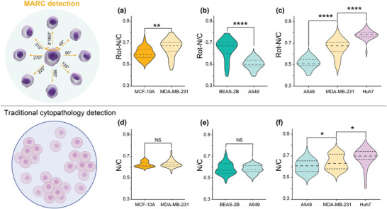
Figure 6.

N/C‐based examination of cells. Rotation‐based N/C comparison between a) breast healthy cell MCF10A and breast cancer cell MDA‐MB‐231, between b) lung healthy cell BEAS‐2B and lung cancer cell A549, and among c) three cancer cells: lung cancer cell A549, breast cancer cell MDA‐MB‐231 and liver cancer cell Huh‐7. Traditional cytopathology detection one angle quantification based N/C comparison between d) breast healthy cell MCF10A and breast cancer cell MDA‐MB‐231, between e) lung healthy cell BEAS‐2B and lung cancer cell A549, and among (f) three cancer cells: lung cancer cell A549, breast cancer cell MDA‐MB‐231 and liver cancer cell Huh‐7. All cell lines were analyzed based on the quantification of 30 cells, per cell averaged by the N/C value of 8 angles. All p‐values were determined using one‐way ANOVA. NS: no significance. * p < 0.05, ** p < 0.01, **** p < 0.0001.
