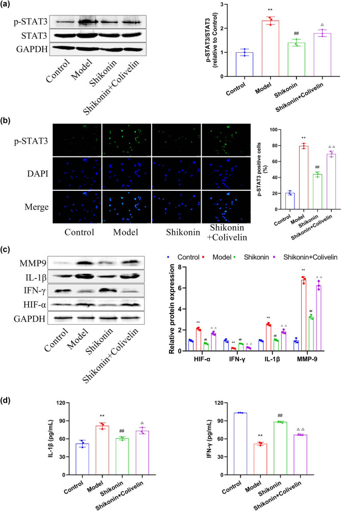
Figure 5.
Activation of STAT3 can reverse the inhibitory effect of shikonin on the function of MLE-12 cells. (a) Western blot analysis was performed to measure the expression levels of STAT3 and p-STAT3 in mouse epithelial cell line MLE-12. (b) The localization of p-STAT3 in mouse bronchial epithelial cell line MLE-12 was detected by immunofluorescence staining. (c) Western blot was applied to measure the expression levels of HIF-α, IFN-γ, IL-1β, and MMP9. (d) ELISA was used to detect the concentrations of inflammatory factors (IL-1β and IFN-γ) in the MLE-12 cell medium. Compared with the control group, ** p < 0.01; compared with the model group, ## p < 0.01; compared with the shikonin group, △ p < 0.05; compared with the shikonin group, △△ p < 0.01, n = 3.

