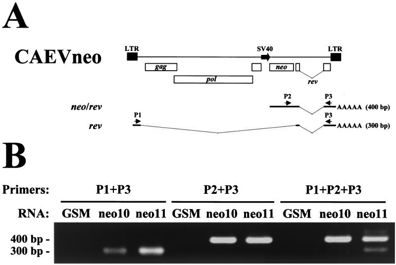
FIG. 3.
Semiquantitative RT-PCR assay of double-spliced rev transcripts in GSM cells infected with CAEVneo10 or CAEVneo11. (A) The CAEVneo10 and CAEVneo11 integrated proviral structure is shown to indicate the relative positions of primers P1, P2, and P3. Thick lines indicate the rev and predicted neo/rev exons. Bent dotted lines indicate introns. The expected sizes of amplicons obtained by RT-PCR for each transcript are shown on the right. (B) Ethidium bromide-stained agarose gel with RT-PCR products. The size of the amplified fragments is indicated on the left. Primers for PCR are indicated above the gel. The RNA used for RT-PCR is indicated above each lane. GSM, uninfected GSM cell RNA; neo10, CAEVneo10-infected GSM cell RNA; neo11, CAEVneo11-infected GSM RNA.

