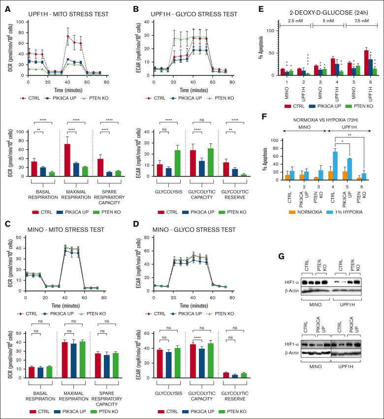
Figure 2.
PTEN KO and PIK3CA UP cell lines have changed the activity of key energy-metabolic pathways and have increased survival under hypoxia. (A-D) Mito stress tests and glycol stress tests performed using Seahorse analyzer. (A) Decrease in the basal respiration, maximal respiration, and spare respiratory capacity in UPF1H PIK3CA UP and PTEN KO cells compared with UPF1H unmodified cell line; measured by OCR. (B) Increase in basal glycolysis in UPF1H PTEN KO cells, decrease of glycolytic capacity in UPF1H PIK3CA UP cells, and decrease of glycolytic reserve in both tested variants; measured by ECAR. (C) No significant changes of mitochondrial function in MINO PIK3CA UP modified cells compared with MINO CTRL. (D) Slight decrease of glycolysis parameters in in MINO PIK3CA UP modified cells compared with MINO CTRL. (E) Number of apoptotic cells 24 hours after exposure to the inhibitor of glycolysis 2-DG (2.5, 5, and 7.5 mM); apoptosis of the treated cells was normalized to the apoptosis of the untreated cells (n = 3). (F) Increased survival of PTEN KO and PIK3CA UP cell lines after 72-hour exposure to 1% hypoxia; apoptosis of the cells cultured for 72 hours under 1% hypoxia (as well as apoptosis of the cells cultured for 72 hours in parallel under normoxia) was normalized to the apoptosis of the cells before placement into hypoxia (n = 3). (G) Western blot analysis of HIF1-alpha in PTEN KO and PIK3CA UP cell lines compared with respective control cell lines (for each sample, n = 2); data are represented by means ± SD; ∗P < .05; ∗∗P < .01; ∗∗∗P < .001; ∗∗∗∗P < .0001; (N) represents the number of biological replicates. CTRl, XXX; ns, not significant.

