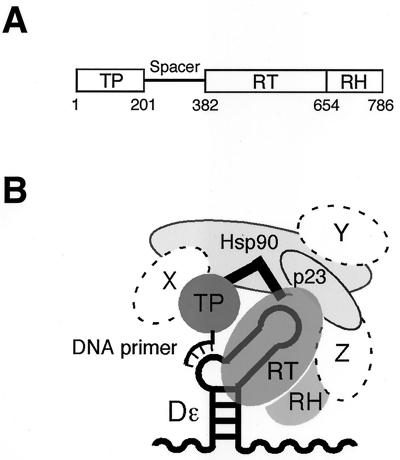
FIG. 1.
(A) Domain organization of hepadnaviral P proteins. Numbers indicate amino acid positions of DHBV P. (B) Model of the DHBV replication initiation complex. P protein with its TP and RT-RH domains connected via the spacer (black angled bar) is bound to the RNA stem-loop Dɛ. A bulged region in Dɛ serves as a template for the synthesis of a short DNA primer that is covalently linked to a Tyr residue in the TP domain. Binding to Dɛ requires P protein to be present in a multicomponent complex composed of Hsp90, p23 (light gray objects), and, most likely, additional, as yet unknown, factors (designated X, Y, and Z).

