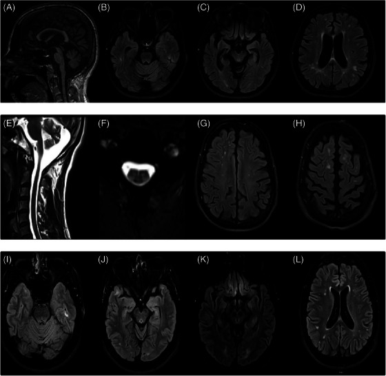
FIGURE 2.

MRI of Case 1. (A) Sagittal T1 sequence showing generalized atrophy and loss of brainstem volume. (B–D) Axial flair sequence showing subcortical and periventricular white matter hyperintensities at three different levels. Magnetic resonance imaging (MRI) of Case 2. (E) Sagittal T1 showing atrophy. (F) Axial T2 images of cervical spinal cord showing posterolateral column hyperintensity. (G,H) Axial fluid‐attenuated inversion recovery (FLAIR) images showing peripheral sulci widening, and subcortical hyperintensities. MRI of Case 3. (I–L) Axial FLAIR images at different levels evidence small white matter hyperintensities and gliosis in the straight gyrus.
