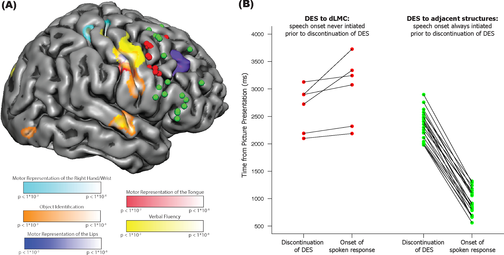
Fig. 1. Stimulation of dorsal Laryngeal Motor Cortex disrupts vocalization during speech production.

(A) Overview of intra-operative direct electrical stimulation mapping and pre-operative functional MRI. Reconstruction of the right hemisphere of Patient AJ’s brain, with locations of intraoperative stimulation associated with errors (red circles) and correct trials (green circles). The tumor in the right frontal lobe is represented in purple (visible in the mesh where it came to the cortical surface). Also plotted on the cortical surface are the results of pre-operative functional MRI for wrist, lip, and tongue movements, object identification, and verbal fluency. (B) Speech production is disrupted by stimulation to dLMC but not to adjacent structures. All data points are temporally aligned (y-axis) with respect to the onset of the picture stimulus on the corresponding trial. The left panel plots data for stimulation to dLMC (red circles) while the right panel plots data for stimulation to structures adjacent to (but not including) dLMC. The left column of data points in each panel corresponds to the time-point, after picture presentation, when direct electrical stimulation to the cortical surface was discontinued; the right column in each panel plots the time point (post picture onset) at which speech initiated (the two measures for each trial connected by line). The key observation is that for trials involving stimulation to dLMC (left panel), it was always the case that onset of a correct response occurred after electrical stimulation was discontinued. By contrast, when stimulation was applied to structures adjacent to dLMC (right panel), the patient was always able to initiate a correct response prior to discontinuation of stimulation. This pattern was substantiated with formal analysis: For those trials in which the patient eventually produced an accurate response after stimulation to dLMC, the average response time (from picture stimulus onset to initiation of a correct response) was 2983ms (SD=605ms). That response time was longer than the average response time for the 21 accurate trials associated with stimulation to structures adjacent to, but not overlapping with, dLMC (Response Time=939ms, SD=229ms; Welch Two Sample t-test, t=8.11, p<.001). Importantly, the bipolar stimulator was not in contact with the brain for a longer period of time on trials of transient speech arrest involving stimulation to dLMC (mean = 2277ms, range = 1800:2740ms) compared to trials with stimulation to surrounding structures (mean = 2103ms, range = 1530:3200ms; t =1.02, p=.33).
