
Figure 1.
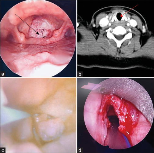
(a) Near-total obstruction of the glottis (black arrow mark showing papilloma). (b) Coronal section of computed tomography scan of the neck at the subglottic level (red arrow showing no subglottic invasion of papilloma). (c) Bronchoscopic image showing guidewire-assisted intubation; (d) Vocal cords after debulking surgery